History of FISH

Fluorescence in situ hybridization (FISH) is a powerful molecular biological technique that allows us to visualize and map the location of specific DNA or RNA sequences in cells and tissues. It has contributed greatly to our understanding of genetics, gene expression, chromosomal abnormalities and various biological processes.

The history of FISH is marked by significant developments and milestones:

1. Early concepts and developments (1960s and 1970s).

  • The foundation for FISH was laid in the 1960s when researchers began experimenting with nucleic acid hybridization techniques. Hybridization involves linking together complementary strands of DNA or RNA.
  • In 1969, John Cairns (pictured) used radioactive probes to detect DNA sequences in the bacterial genome, which was one of the first instances of in situ hybridization.
  • In the early 1970s, researchers such as Mary-Lou Pardue and Joseph Gall used radioactive RNA probes to visualize specific gene sequences on Drosophila (fruit fly) chromosomes.

2. Introduction of non-radioactive probes (1980s).

  • The use of radioactive probes led to concerns about user safety and disposal of the material, which led to the development of non-radioactive probes for FISH.
  • In 1980, Louise Brown and John Smith published a method using biotin-labeled probes for in situ hybridization.
  • Throughout the 1980s, researchers explored alternative labeling techniques, such as digoxigenin-labeled probes, which produced detectable signals without the use of radioactivity.

3. Fluorescent-labeling and confocal microscopy (1980s and 1990s)

  • The breakthrough for FISH came with the integration of fluorescent dyes as labels for FISH probes. This enabled easier detection and visualization of hybridization events.
  • In 1986, John W. Sedat and David A. Agard introduced wide-field epifluorescence microscopy to image FISH probes.
  • In 1989, Robert Singer and colleagues developed the method of fluorescence in situ hybridization using directly labeled DNA probes.

4. Chromosome painting and spectral karyotyping (SKY) (1990s-2000s).

  • Researchers began using multiple fluorescent probes to simultaneously label different chromosomes, a technique known as chromosome painting.
  • In 1995, the Spectral Karyotyping (SKY) technique was introduced, which allowed the simultaneous visualization of all chromosomes using different fluorescent colors.
  • These techniques revolutionized research into chromosomal abnormalities and genetic variations.
  •  

5. Advances in probe design and automation (2000-present).

  • FISH has found applications in cancer diagnostics, prenatal testing and several other research areas.
  • Biotrack(NL-Lab's parent company) is developing C-FISH, a patented automated version of FISH that uses powerful computers, high-resolution cameras and AI-driven algorithms.
  • The Biotrack platform, based on C-FISH technology, enables efficient analysis of large numbers of samples, including for complex applications (such as gut microbiome research).
  • Biotrack is continuously working on innovations in probe design, fluorescent-labeling technologies, digital image processing and AI-driven algorithms to further improve the unprecedented sensitivity, specificity and speed of FISH.

The history of FISH illustrates the evolution of molecular techniques and their profound impact on our understanding of genetics and biology. FISH remains a fundamental tool for studying genome organization, gene expression and chromosomal aberrations in various organisms and cell types.

Interested or questions?

If you have any questions as a result of this article, we warmly encourage you to contact us.

Sharing:

Also read...

The time between the collection of a (stool) sample and its processing in a laboratory has a...

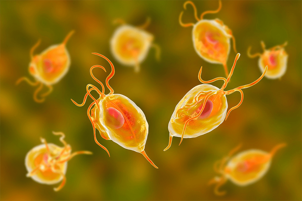
Many people think that parasites are only found in distant lands, but nothing could be further from the truth. In this article...

In our view, the classical dysbiosis model is too limited and outdated because it works with only two different...